Address
93/D LIC Colony, Tagore Town.
Prayagraj 211002
Work Hours
Monday to Friday: 11AM - 5PM
Address
93/D LIC Colony, Tagore Town.
Prayagraj 211002
Work Hours
Monday to Friday: 11AM - 5PM
Use of rotational atherectomy and hybrid approach to treat multiple stenosis and in-stent restenosis
Abstract
In-stent restenosis (ISR) rising in bare-metal stents (BMS) and drug-eluting stents (DES) is challenging to treat. We present the case of a 61-year-old male patient who had triple vessel disease, ISR, and pre-existing coronary artery disease (CAD). The optimal lesion preparation strategies, such as rotational atherectomy and balloon angioplasties, were required due to severe stenosis and ISR. Successful percutaneous transluminal coronary angioplasty (PTCA) was performed using a hybrid approach including DES and bioresorbable scaffold (BRS). Optical coherence tomography(OCT) revealed the successful deployment of BRS with firmly embedded struts within the vessel wall without malappositionand edge dissection.
Introduction
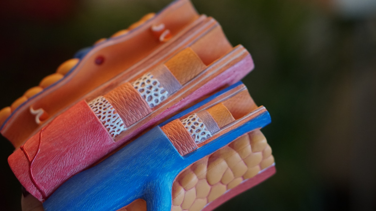
In-stent-restenosis (ISR) is characterized by the presence of >50% lumen diameter stenosis at the previously implanted stent’s site or its 5-mm edges. In the pre-stent period, the prevalence of restenosis ranged from 32-55% of all angioplasties, which was reduced to 17-41% with the use of BMS. The advent of drug-coated balloons (DCB), DES, and BRS has brought down the restenosis rate to less than 10% (1). In ISR cases, the revascularization strategy requires adequate lesion preparation to further reduce the risk of restenosis and enhance the efficacy of DES and BRS. The techniques for preparing lesions include rotational atherectomy, cutting balloons, semi-compliant and non-compliant balloon angioplasty, and, more recently, intravascular lithotripsy (2). Rotational atherectomy is used in <5% of percutaneous coronary interventions (PCI) to treat ostial, severely stenosed, and calcified lesions that are too severe to enable the delivery of a stent to the target site. It eliminates atheromatous plaque by differential cutting, which involves scraping away at the stiff calcified plaque with microscopic (20 to 50 µm) diamond chips affixed to the surface of an olive-shaped burr rotating at a high speed (150,000 to 200,000 rpm). Rotational atherectomy eliminates superficial calcification to increase lesion compliance before balloon dilatation and stent implantation. It is effective only in calcified and inelastic lesions, unlike soft and thrombus-containing lesions(3).
Implantation of coronary stents during PTCA is a preferred interventional therapeutic method to restore normal blood flow through stenotic coronary arteries. It is proven that using DES instead of BMS lowered the risk of restenosis, however, treating ISR driven by neointimal proliferation/hyperplasia or calcification remains a challenging question for interventional cardiologists. The aim of developing BRS was to provide short-term benefits of permanent stents with the added advantage of completely degrading over 2-3 years. This allows full recovery of the vasomotor and endothelial function of the target vessel. This “leave nothing behind” strategy of BRS prevents long-term inflammation, preserves distal bypass grafting sites, and allows unimpeded future vessel imaging (4). To the best of our knowledge, this is the only case in which rotational atherectomy and a BRS implant were combined to treat ISR.
Case presentation
A 61-year-old male patient presented with complaints of radiating pain from left shoulder to left hand followed by a tingling sensation while walking for 3-4 months. The patient was hypertensive, non-diabetic, non-smoker, and non-alcoholic without a history of substance/drug abuse and allergies, he had a history of renal disease in 1995. He was also a known case of CAD and underwent PTCA to obtuse marginal 1 (OM1) and mid-left anterior descending artery (LAD) with Endeavor DESs (Medtronic’s, Minneapolis, USA) (3.5×18 mm and 3×24 mm) size respectively in 2008. Post-PTCA, he was diagnosed with dyslipidemia. On 26 August 2022, the patient was admitted for further evaluation and management of CAD. On general examination, the patient was found to be conscious, cooperative, and oriented to time, place, and person. Upon physical examination, the patient was found to be afebrile, with a soft abdomen, a respiratory rate of 20 breaths/minute, and normal S1 and S2 levels with bilateral equal air entry. His vital signs showed a heart rate of 78 beats/min and blood pressure of 140/80 mmHg. His biochemical tests revealed normal levels of LDL-cholesterol (7.8 mg/dl), triglycerides (138 mg/dl), HbA1c (5.6%), and serum creatinine (1.23mg/dl). Radiological examination showed normal X-ray and no significant changes in electrocardiography. 2D echocardiography revealed no regional wall motion abnormalities and normal left ventricular function with an ejection fraction of 60%. Right femoral approach angiography revealed triple vessel disease with 95-99% stenosis in the left circumflex coronary artery to obtuse marginal 1 (LCx-OM1)(Figure 1A) and ~ 95% stenosis in the mid-LADdue to ISR followed by 70-80% stenosis in diagonal 2 (D2) (Figure 1B). In addition, it was found that the mid-right coronary artery (RCA) had a ~70-80% stenosis, RCA-posterior left ventricular artery(R-PLV) had 90% stenosis, and distal-RCA had 70% stenosis (Figure 1C).
| D |
Figure 1: (A) Diagnostic angiogram showing 90-99% subtotal occlusion in LCx-OM1 due to ISR (B) ~ 95% stenosis in mid-LAD due to ISR. (C) Stenosis in mid-RCA (70-80%), 90% R-PLV followed by 70% in distal. (D) Rotablation using RotaLink Plus 1.50mm (0.059″).
Revascularization using a hybrid technique
In the first step of the procedure, the right femoral approach was adopted using a 4F Micropuncture kit, followed by the insertion of a 7F femoral sheath and 6F Cordis XB 4.0 guiding catheter to treat ISR in LCx-OM1. The procedure was then initiated using Runthroughguidewire (0.014″x180 cm) (TerumoInterventional Systems, Tokyo, Japan). However the lesion in the LCx-OM1 was highly stenosed and calcified, another guidewire ASAHI PTCA guidewire Feilder (0.014″x180 cm) was used. After subsequent failure, the lesion was finally crossed using a more supportive microcatheter, high penetration chronic total occlusion ASAHI Gaia-Second (0.014″x190 cm) guidewire. This was exchanged with ROTAwire Floppy (0.009″x330 cm) with the aid of a microcatheter. Further, the RotaLink Plus (0.059″x1.50 mm) (Boston Scientific, Massachusetts, USA) rotablator burr was required to shave off the accumulated, hardened, and calcific plaque in the LCx-OM1 vessel (Figure 1D). Following rotablation, the first pre-dilatation was performed using an NC Trek balloon (3.0×20 mm at 16-20 atm for 10 sec) (Figure 2A) and PTCA Angiosculpt scoring balloon (3.5×15 mm at 14-18 atm for 10 sec) was required for second pre-dilatation (Figure 2B) for adequate lesion preparation. Angioplasty was performed by implanting XIENCE Alpine DES(3.5×28 mm at 12 atm for 15 sec) (Abbott Vascular, CA, USA) (Figure 2C) followed by a successful post-dilatation with Lineage NC balloon (3.5×15 mm at 16-20 atm for 10 sec) as shown in Figure 2D.
| B |
Figure 2: (A) Pre-dilatation using NC Trek balloon (3.0×20 mm at 16-20 atm for 10 sec) and (B) Angiosculpt PTCA balloon (3.5x15mm at 14-18 atm for 10 sec). (C) Stent deployment Xience Alpine (AbbottVascular, CA, USA) (3.5x28mm at 12 atm for 15 sec). (D) Post-dilatation using Lineage NC balloon (3.5x15mm at 16-20 atm for 10 sec).
To treat ISR in the mid-LAD, the lesion was accessed using 6FCordis XB 4.0 catheter. Initially, a workhorse guidewire (Runthrough 0.014″x180cm)(TerumoInterventional Systems, Tokyo, Japan) was used to cross the lesion, but this was unsuccessful. Therefore, ASAHI PTCA guidewire Feilder (0.014″x180 cm) was employed due to tight stenosis and lesion crossing was achieved. Pre-dilatations were performed thrice using balloons of ascending diameters for optimal bed preparation. The initial pre-dilatation was carried out using NC Trek (2×15 mm at 20-24 atm for 10 sec), followed by NC Trek (3×20 mm at 20-24 atm for 10 sec), and finally using an Angiosculpt PTCA scoring balloon (3.5x15mm at 14-16 atm for 10 sec)(Figure 3A). LAD-D2 with 70-80% stenosis was treated with balloon angioplasty only using an NC Trek balloon (2 x 15 mm balloon at 14-16 atm for 10 sec)(Figure 3B). After pre-dilatation in mid-LAD, MeRes100 BRS (Meril Lifesciences, Vapi, India) (3.5×29 mm at 4-10 atm for 15 sec) (Figure 3C) was deployed at the site of occlusion to revamp the blood flow. Lastly, post-dilatation was performed using an NC Trek balloon (3.5×15 mm at 20-24 atm for 10 sec)(Figure 3D).
Figure 3: (A) Pre-dilatation in mid-LAD using Angiosculpt PTCA scoring balloon (3.5x15mm balloon at 14-16 atm for 10 sec). (B) Balloon angioplasty in LAD D2 using NC Trek balloon (2x15mm at 14-16 atm for 10 sec). (C) BRS MeRes100 deployment (3.5x29mm at 4-10 atm for 15 sec). (D) Post-dilatation using NC Trek balloon (3.5x15mm balloon at 20-24 atm for 10 sec).
Figure 4: (A) Final angiogram and (B) successful deployment of BRS MeRes100 (Meril Lifesciences) in mid-LAD as observed under OCT.
Discussion
With the rising incidence of stent implantation, ISR has become a challenge in interventional cardiology. Neointimal proliferation and calcium deposition at the stent lumen cause the recurrence of ISR. The potential treatment strategies required are balloon angioplasty, rotational atherectomy, DCB, DES, and most recently BRS deployment. The most efficacious treatment strategy depends on the patient and the lesion characteristics. Recent developments in DCB and DES with antiproliferative drugs have significantly reduced ISR incidences (1). BRS provides functions of DES with the additional benefit of bio-resorbability. BRS such as (MeRes-100 Meril Lifesciences) is a next-generation sirolimus-eluting bioresorbable scaffold with a strut thickness of 100μm. It has a lower crossing profile for easy delivery and early endothelialization. Intravascular imaging techniques such as OCT and intravascular ultrasound (IVUS) help in understanding the pathophysiology of ISR and aid in optimal stent deployment without malapposition (5).
This case presents the ISR in LCx-OM1 and mid-LAD of the patient who has previously been stented with Endeavor DESs (Medtronic’s, Minneapolis, USA)in 2008. Depending on the lesion severity, different revascularization techniques were employed for optimal lesion preparation, to lower risks, and to improve clinical outcomes. Multiple guidewires such as Runthrough, Feilder, and Gaia-Secondwere used to cross the lesion. Since the lesion in the LCx-OM1 was long, diffused, un-dilatable, and stiff due to excessive calcium deposition or neointimal hyperplasia, it mandated the use of a rotablator —“RotaLink Plus” (0.059″x1.50mm) (Boston Scientific, Massachusetts, USA). It is a safe and effective technique if performed by professional operators to treat hard calcific plaque. The safety and efficacy of this device in the treatment of calcific lesions/ISR has previously been established (6–7). The rotating burr at the tip of the rotablator assisted in removing the buildup of hardened plaque in OM1 of the LCx vessel and aided in dislodging the coronary blockage. Post rotablation, LCx-OM1 underwent a successful deployment using DESXIENCE Alpine (Abbott Vascular, CA, USA). The patient had CAD, hypertension, dyslipidemia, and surgical risk factors, which are considered prognostic factors for long-term PCI outcomes. Considering these factors, to minimize the metallic load and to reduce the risk of repeat revascularization BRS (MeRes-100 Meril Lifesciences, Vapi, India) was employed to treat mid-LAD ISR. BRS offers enhanced relief from late ST and ISR by completely dissolving after arterial healing, restoring normal vasomotion, and without any stent material residues within 2-3 years. Lastly, 70-80% of the lesion in LAD D2 was also successfully treated with balloon angioplasty.
Conclusion
The hybrid strategy should be considered for PCI conducted for long calcific lesions and ISR cases, particularly in complex cases involving multi-vessel disease. The critical factors to consider for treatment planning include the selection and crossability of the guidewire through the plaque, reducing the ischemic burden of the coronaries, and the accurate treatment strategy to ensure a successful intervention ASAHI Gaia-Second guidewire should be considered when attempts through more conventional guidewires are unsuccessful. Rotablation atherectomy is needed when severely calcific coronary plaques are encountered. The MeRes100 BRS is a sirolimus-eluting thin-strut scaffold that may be recommended for the subset of high-risk patients with ISR having cardiac risk factors to reduce the need for further revascularization. Using OCT guidance, interventionalists may assure accurate strut coverage without malapposition and improved myocardial perfusion following the procedure.
For the publication of this case report, the patient’s consent was taken. The identity of the patient has not been disclosed.
No conflict of interest.
Each author has significantly contributed to the idea and design of the study, the acquisition of data, analysis and interpretation of data, writing of the paper, its critical revision for key intellectual content, and its final approval for submission.
The authors would like to acknowledge the patient’s family for their consent.
Abbreviations
BMS Bare-metal stents
BRS Bioresorbable scaffold
CAD Coronary artery disease
D2 Diagonal 2
DCB Drug-coated balloons
DES Drug-eluting stents
ISR In-stent restenosis
IVUS Intravascular ultrasound
LAD Left anterior descending artery
LCx-OM1 Left circumflex coronary artery to obtuse marginal 1
OM1 Obtuse marginal 1
OCT Optical coherence tomography
PCI Percutaneous coronary interventions
PTCA Percutaneous transluminal coronary angioplasty
RCA Right coronary artery
R-PLV RCA-posterior left ventricular artery
References
1. Buccheri D, Piraino D, Andolina G, Cortese B. Understanding and managing in-stent restenosis: A review of clinical data, from pathogenesis to treatment. J Thorac Dis. 2016;8(10):E1150–62. Doi:10.21037/jtd.2016.10.93
2. Gupta T, Weinreich M, Greenberg M, Colombo A, Latib A. Rotational atherectomy: A contemporary appraisal. Interv Cardiol Rev Res Resour. 2019;14(3):182–9. Doi: 10.15420/icr.2019.17.R1
3. Valdes PJ, Nagalli S, Diaz MA. Rotational Atherectomy. 2022 Jul 4. In: StatPearls [Internet]. Treasure Island (FL): StatPearls Publishing; 2022 Jan–. PMID: 29763091.
4. Serruys PW, Garcia-Garcia HM, Onuma Y. From metallic cages to transient bioresorbable scaffolds: Change in paradigm of coronary revascularization in the upcoming decade? Eur Heart J. 2012;33(1):16–25. Doi: 10.1093/eurheartj/ehr384.
5. Madanchi M, Cioffi GM, Attinger-Toller A,Wolfrum M, Moccetti F, Seiler T, Vercelli L, BurkartP,Toggweiler S, Kobza R, Bossard M, Cuculi F.Long-term outcomes after treatment of in-stent restenosis using the Absorb everolimus-eluting bioresorbable scaffold. Open Heart 2021;8:e001776. Doi: 10.1136/openhrt-2021-001776
6. Alfonso F, Coughlan JJ, Giacoppo D, Kastrati A, Byrne RA. Management of in-stent restenosis. Vol. 18, EuroIntervention. Europa Group; 2022. p. E103–23. Doi: 10.4244/EIJ-D-21-01034
7. Sharma SK, Kini A, Mehran R, Lansky A, Kobayashi Y, Marmur JD. Randomized trial of Rotational Atherectomy Versus Balloon Angioplasty for Diffuse In-stent Restenosis (ROSTER). Am Heart J. 2004;147(1):16–22.Doi.org/10.1016/j.ahj.2003.07.002
8. Abdel-Wahab M, Toelg R, Byrne RA, Geist V, El-Mawardy M, Allali A, et al. High-speed rotational atherectomy versus modified balloons prior to drug-eluting stent implantation in severely calcified coronary lesions: The randomized prepare-CALC trial. Circ Cardiovasc Interv. 2018;11(10):1–12. Doi: 10.1161/circinterventions.118.007415